Problem
We inherited the brief as a smart thermometer. The premise, set before we joined, was that better fever measurement would improve malaria diagnosis in rural Uganda. Vinay and I went to Uganda for three weeks to understand that before designing for it.
The thermometer was answering a question nobody was asking. A nurse running a Health Centre II does not need a device to tell him a child has a fever. He puts a hand on a forehead. What he cannot tell is why the child has one.
Malaria is common enough that a fever is usually treated as malaria. Rapid diagnostic tests exist, but they return enough false positives that people have stopped believing them, so the tablets get prescribed either way, just in case. Over-prescribing wears the medicine out. When someone does eventually have malaria, it works less well.
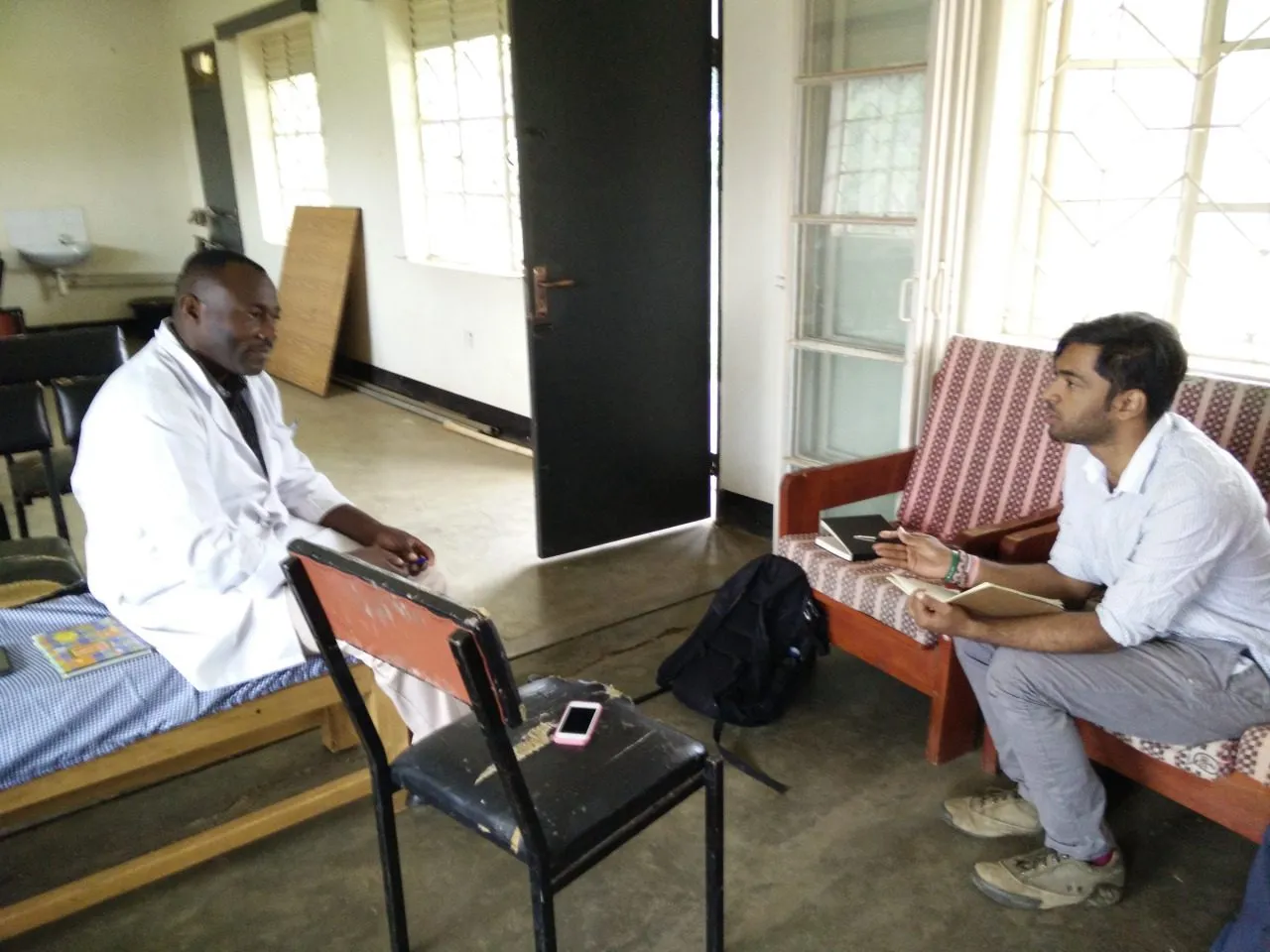
The reliable answer is to look at a blood smear under a microscope and find the parasite. That takes training the people running village health centres do not have. The nurse in charge of one Health Centre II told us he could operate a microscope. What he needed was more microscopes, and more people. He walks about ten kilometres to work because the road will not take a vehicle, and he sees roughly fifty patients, most of them before noon.
Process
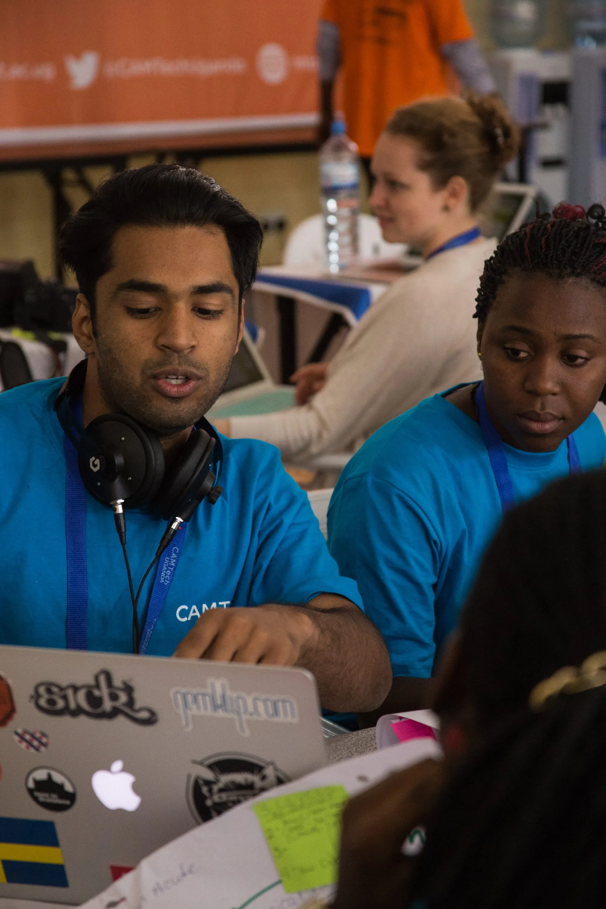
We started at a healthcare hackathon run by CAMTech Uganda, mostly as a way to meet people. It worked. The contacts we made there got us into clinics.
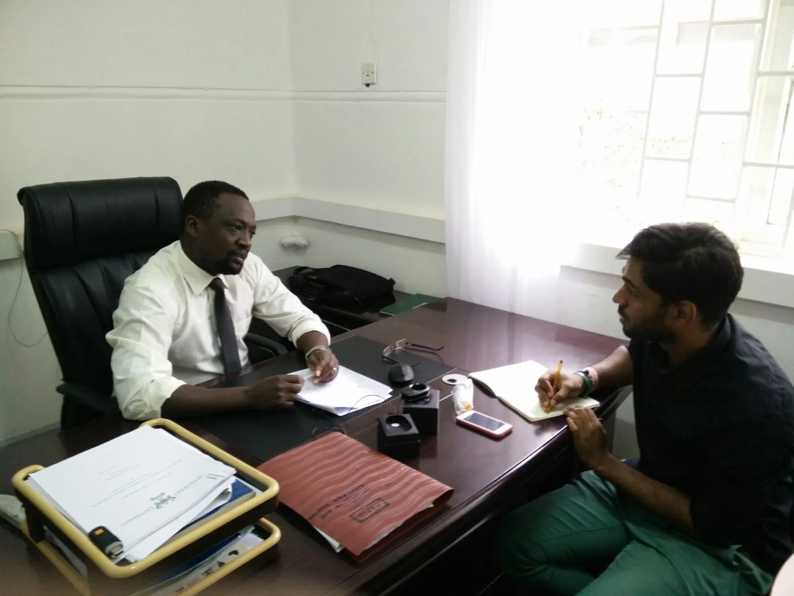
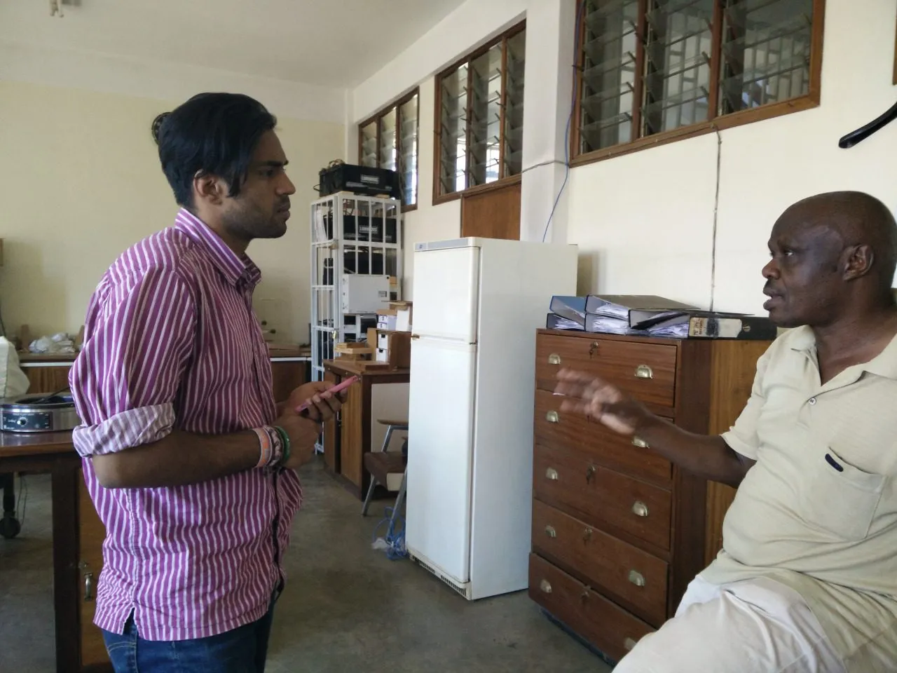
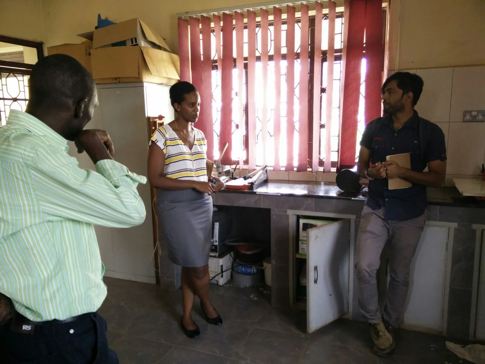
After that we interviewed our way up and down the system: village health teams, health centres at levels two, three and four, a regional referral hospital, a pathologist, a lab technician, a traditional healer, an AI researcher in Kampala, a small-scale manufacturer.
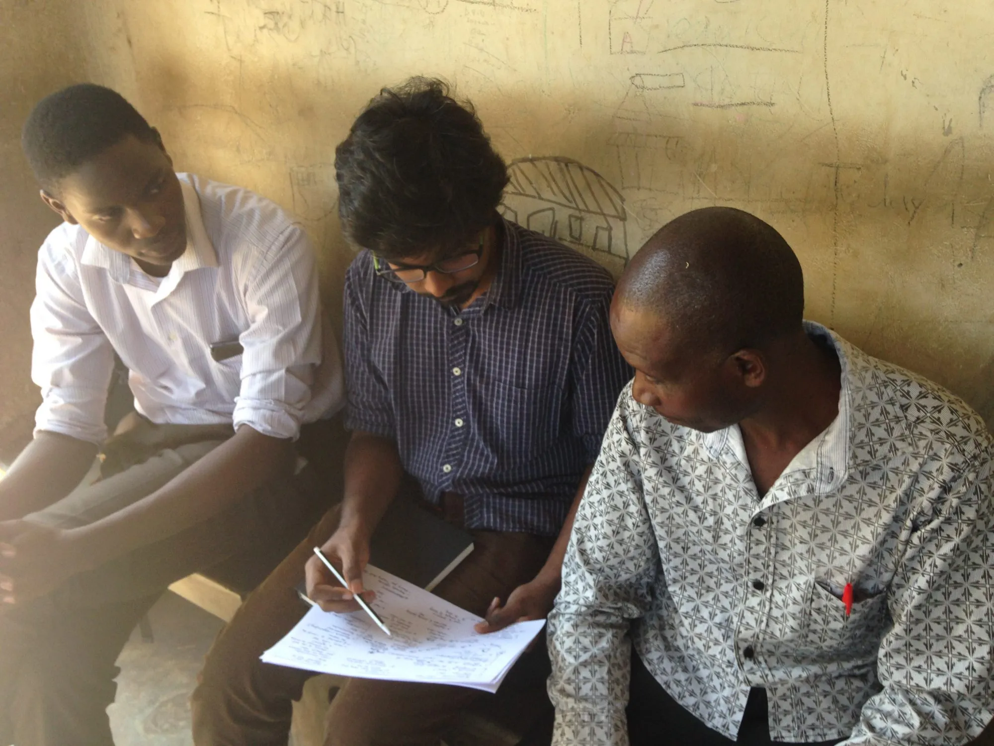
With the village health teams we used a mapping exercise, drawing what happens to a patient one step at a time, then walking back through it with them to check we had it right. It caught things a straight interview did not. People describe the system they are supposed to work in, and then when you draw it, they point at the gaps.
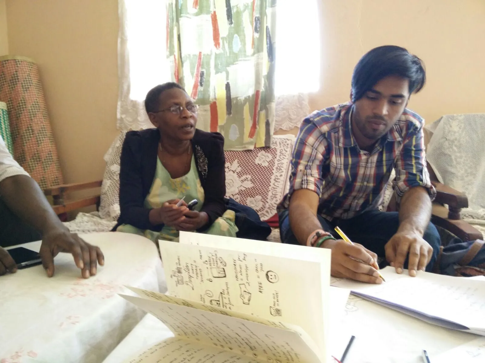
We also spent time on the equipment rather than the people. What arrives, what works, what has been sitting broken for two years because nobody is appointed to calibrate it.
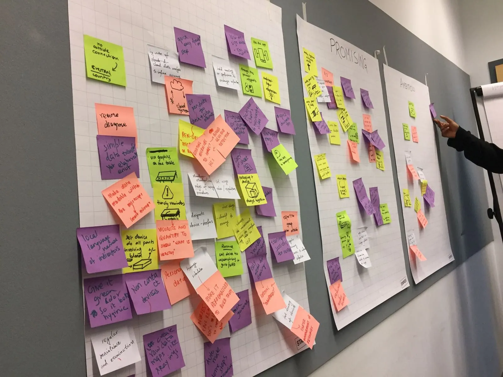
Back in Delft we drew the whole system as one map and marked every problem on it, from a patient with no health literacy on the left to the national medicine supplier on the right.
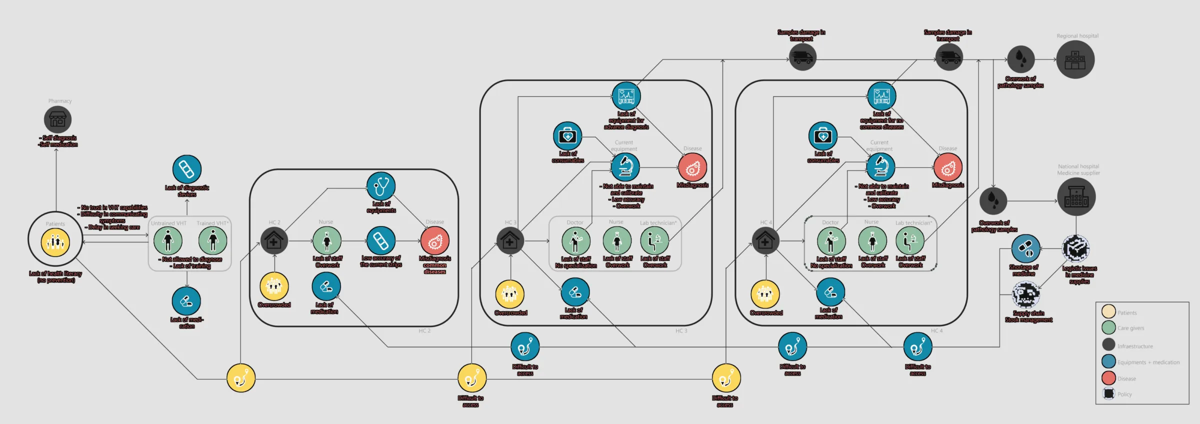
The map made the choice for us. Almost everything a designer could act on sat in Health Centres II, III and IV. Above that, the answer is policy or medicine.
We then argued for a while about whether to aim at Health Centre II or at three and four. Two is harder: less training, nobody to maintain or calibrate equipment, a real risk that whatever we made would not be usable. We took it anyway, because there are far more Health Centre IIs, and because something built for two can be moved up later. The other direction does not work.
The brief we wrote when we got back does not mention thermometers.
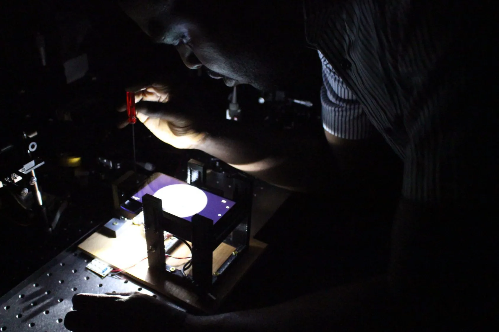
What we proposed instead was to put the microscope where the nurse already is. We took that to the optical engineering group at TU Delft and built it with them, alongside PhD researchers who knew optics far better than we did.
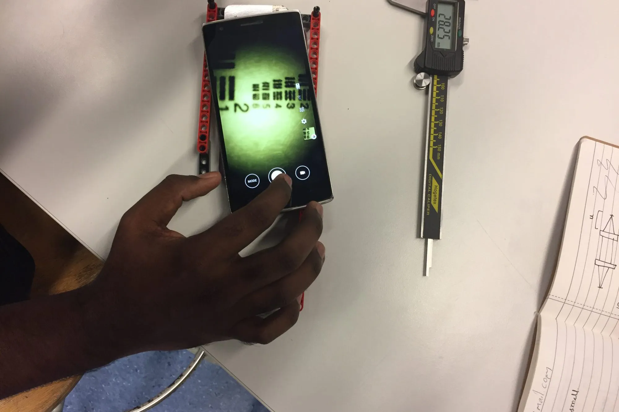
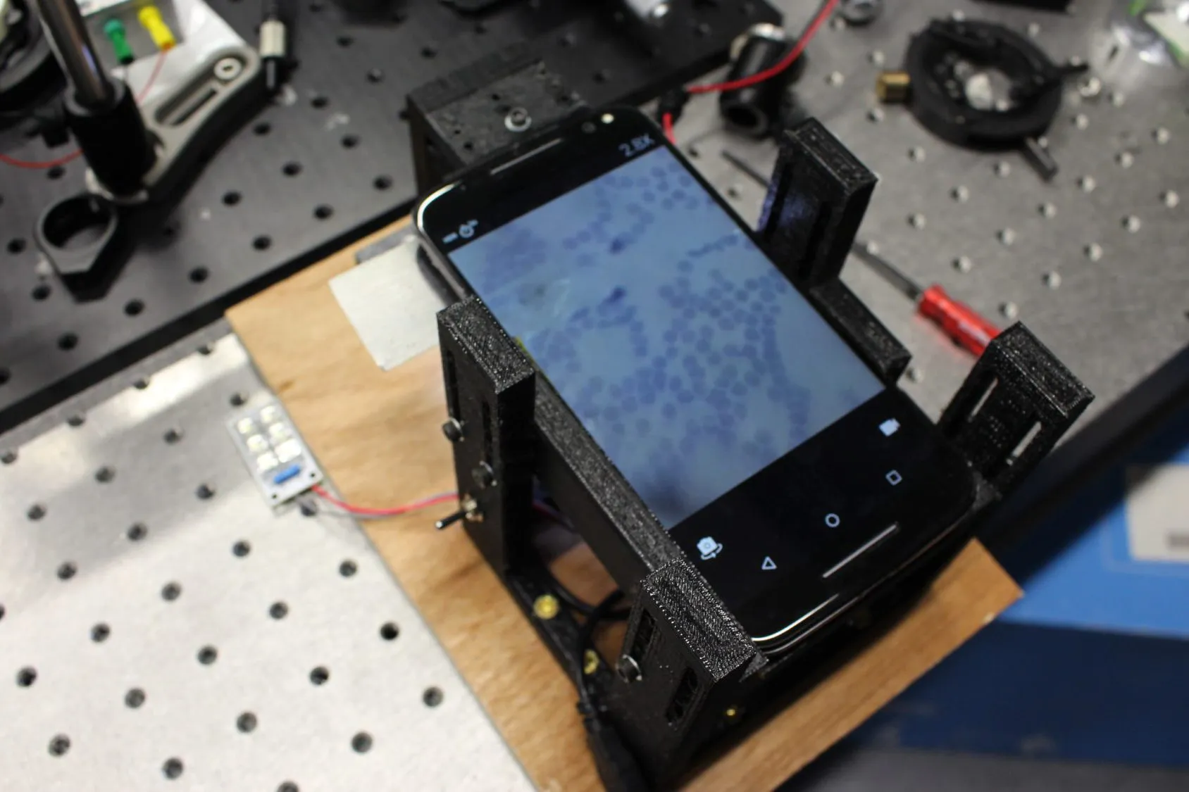
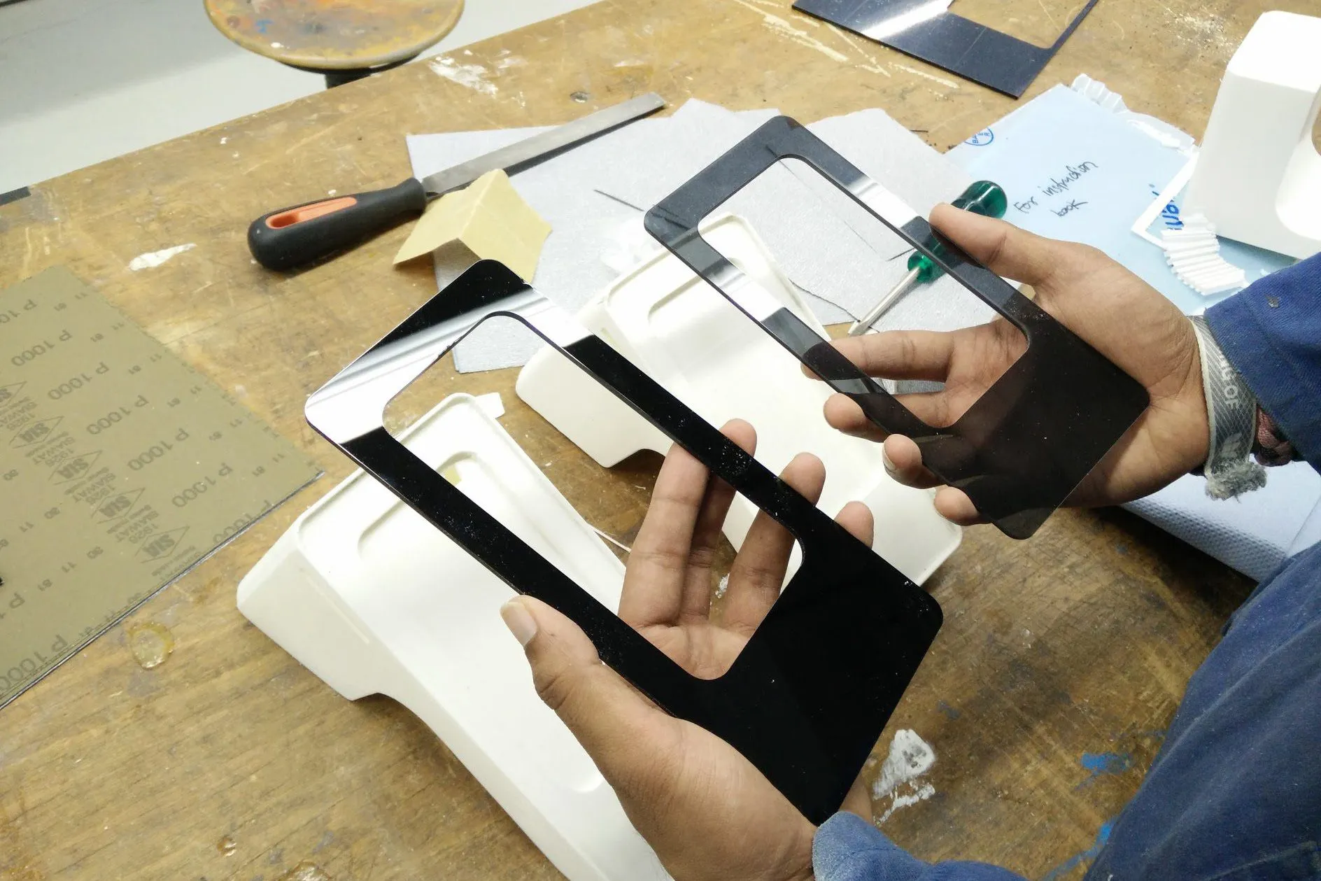
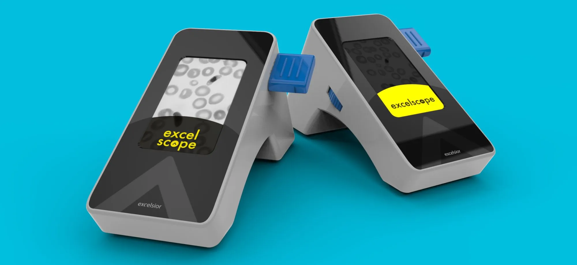
The rule we set was no bespoke electronics. A device with custom parts is expensive to buy and impossible to repair in a village. A phone is already there, already has a camera, a processor and a screen, and can be replaced by anyone. So the design became a body that holds a phone at the right distance above a stack of lenses, an LED array underneath, and a tray for the slide.
Solution
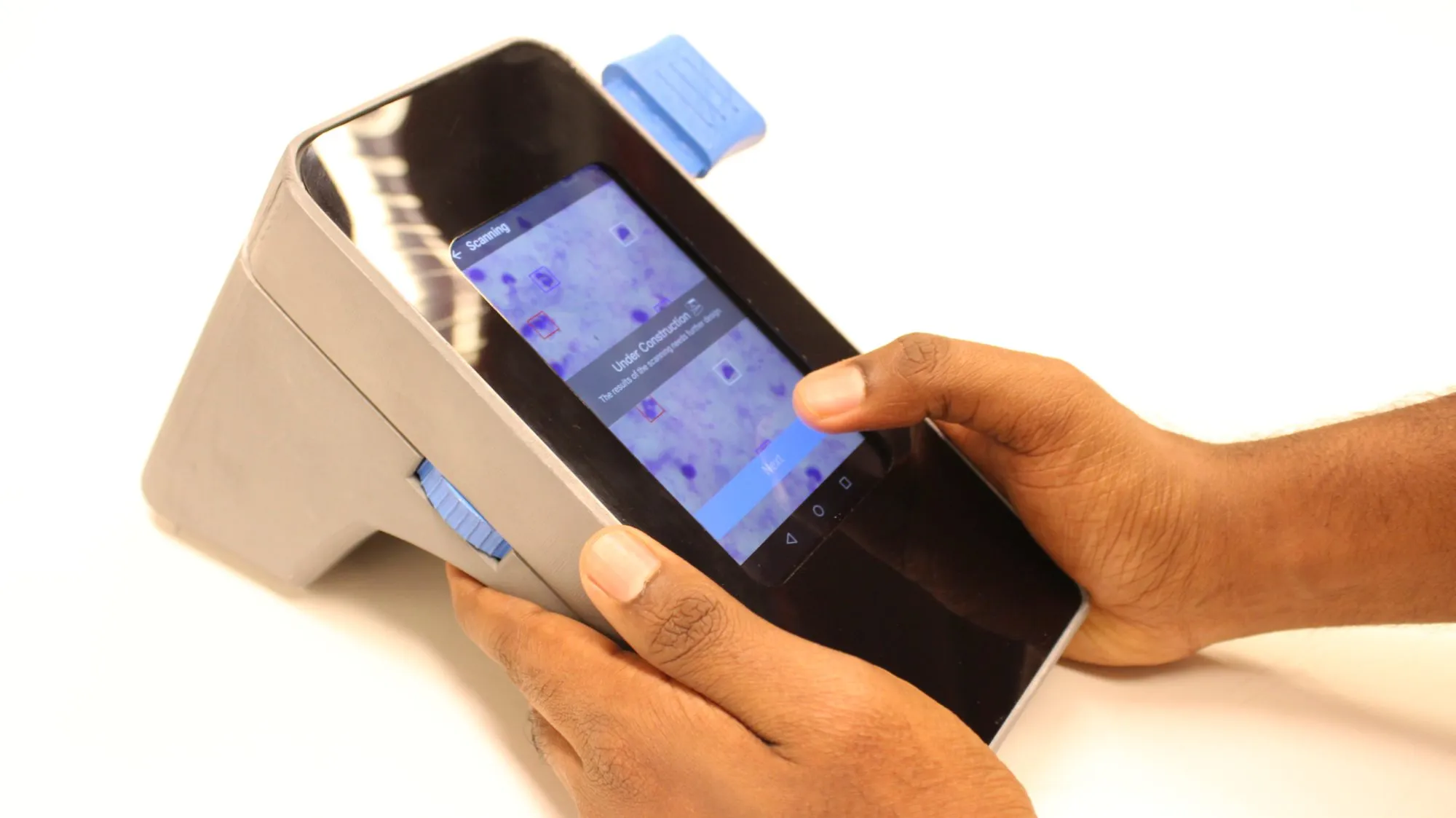
The health worker does the one part that still needs a person: prick a finger and lay a smear on a slide. The slide goes into the tray. The device captures images across it, and the software counts what it finds.
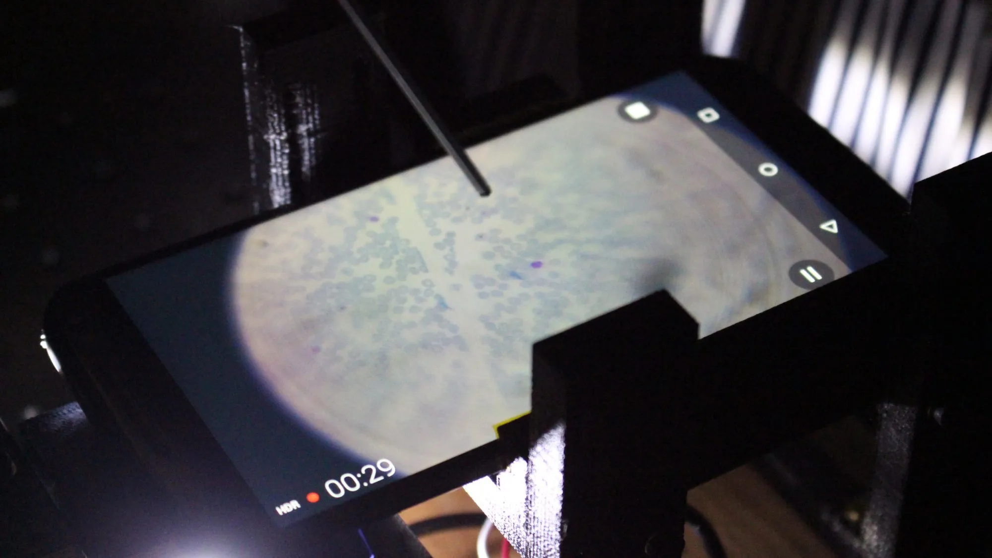
It was not as good as a trained microscopist. In 2016 it was never going to be. It was better than a rapid test, which was the bar that mattered.
When the software is not sure, the images go over the phone network to a doctor elsewhere, who looks at the same slide and answers. Nobody travels, and the slide does not have to survive a bus.
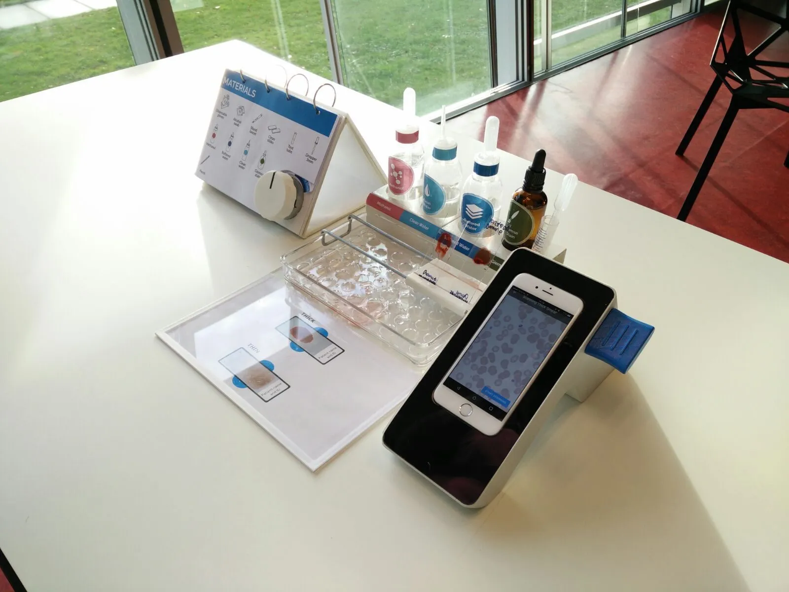
The device on its own would have been useless, though, and that took us a while to accept. Every result depends on the quality of the smear, and the smear is made by someone with no laboratory training. So the thing we actually delivered was a kit: methanol, buffered water, clean water and Giemsa stain in labelled dropper bottles, a staining tray, a laminated card showing what a good thin and thick smear look like with the patient’s name and number written on the glass, and a flip book listing every material with a picture of it. The app is told which kind of smear it is scanning.
Impact
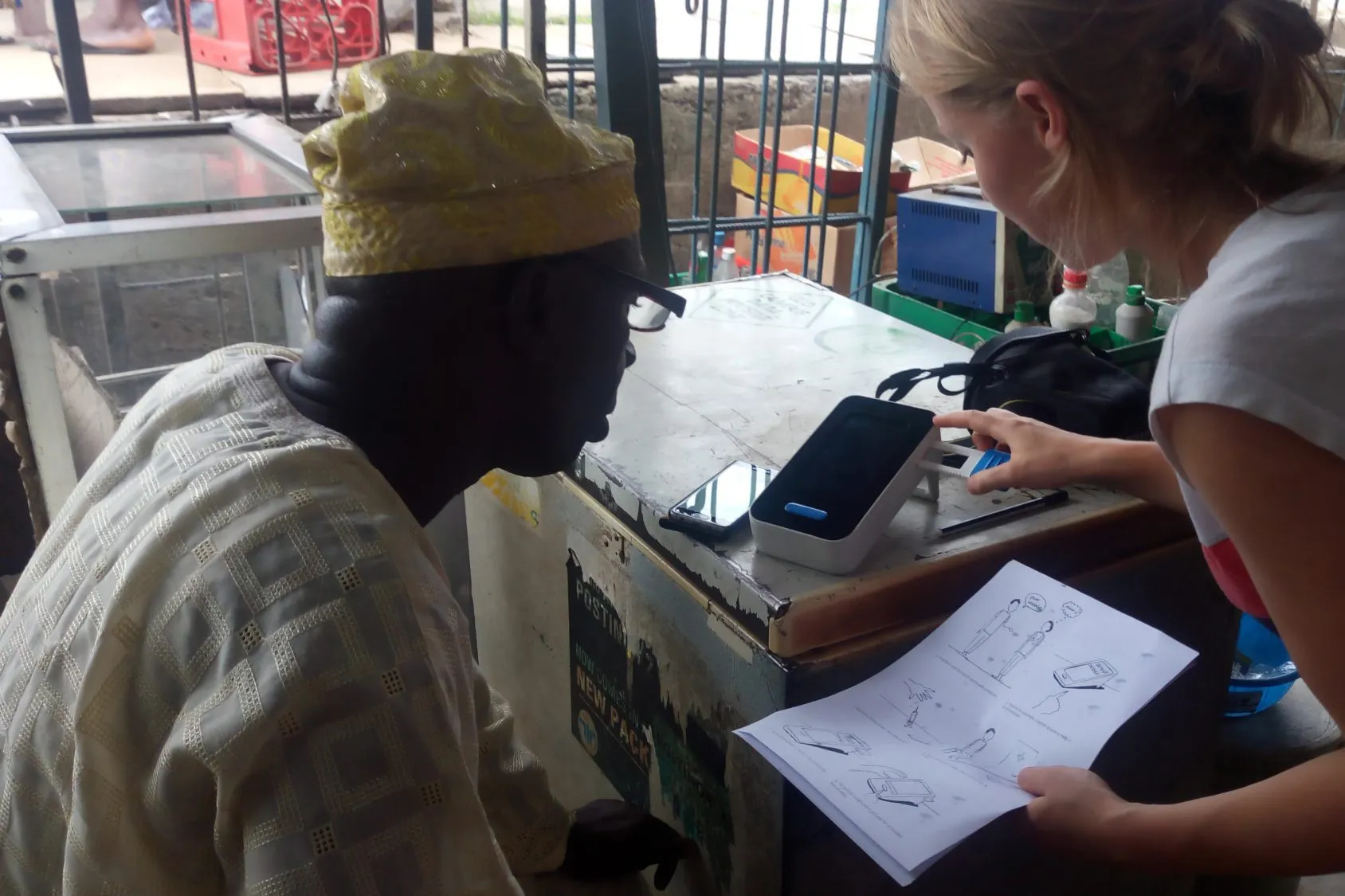
Then it went back. Sitting on a shop counter, being handed to people, being explained with the same illustrated sheets we had drawn.
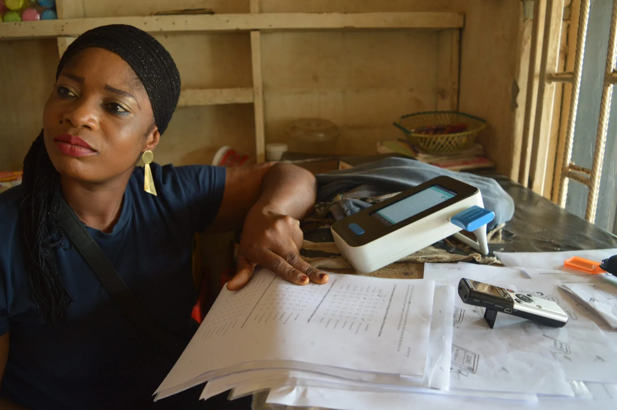
The project outlived us. When our semester ended the optical engineering group took it on and it became a flagship project of the department, carried forward by other students and researchers.
It is now in use in Nigeria, Uganda and other parts of Africa, doing schistosomiasis screening, with malaria in clinical trials.
The part I am most pleased with is not the microscope. It is that we came back and said the thing we had been asked to build should not be built. It cost us the brief we started with, and it was the only honest thing to do with what we had seen.